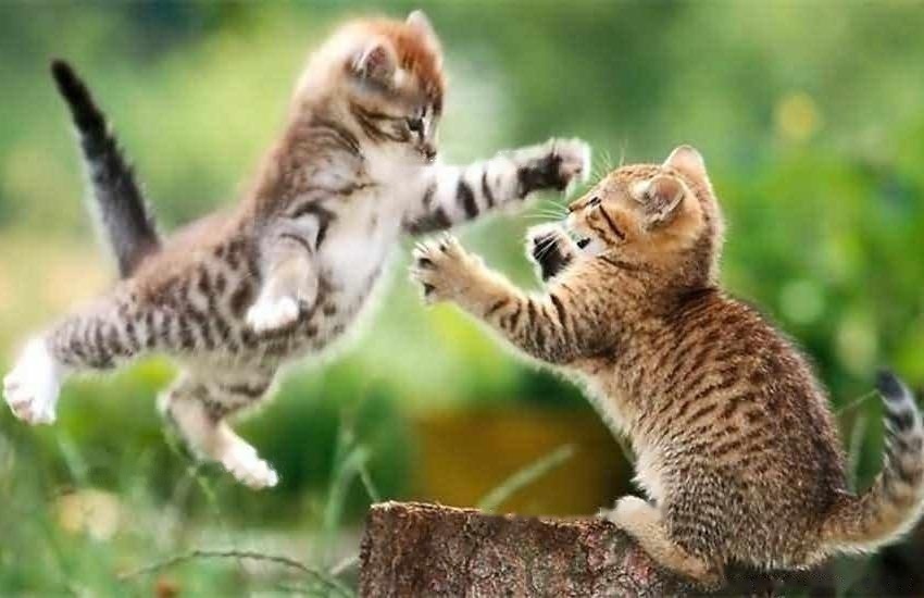
Position : Home / Information

Causes and solutions of pet tear stains

2021-01-23 / 645 Read

Pets's lacrimal gland is an important accessory organ of the eye. Its main function is to secrete tears to moisten the eyeball. Under normal circumstances, the lacrimal gland secretes tears to moisten the eyeball and then enter the nasal cavity through the tear duct, and will not flow out from the corner of the eye. If the lacrimal gland secretes too much tear fluid for some reason and cannot be discharged from the nasolacrimal duct, tears will flow from the corner of the eye, and over time, the hair wetted by excessive tears will leave a brown tear stain [1]. Tears not only affect the facial appearance of pets, but severe and continuous tearing will also become a breeding ground for a series of bacteria, fungi, and fleas, causing eye infections and inflammation, causing pets physical pain.

height=1136

1 Reasons for pet tear marks

1.1 Diet

With the improvement of living standards, pet food variety Different brands of pet food have different nutritional components, and suitable groups of pet dogs and cats are also different. Additives and grains in pet food may cause allergic symptoms, which indirectly lead to increased tear secretion. Too many tears spill out of the eye socket, and the tears contain lactoferrin, which is easily oxidized to form a reddish-brown color, which looks like a rusty color in appearance, which affects the appearance of pets.

1.2 Physiological structure

The dog's eyes are tearing in the wind. Once the lacrimal gland is blocked and inflamed, it is easy to form tear marks. There are some breeds of pet dogs, especially those with short noses. These types of dogs have a large curvature of the nasolacrimal duct. Even the normally secreted tears are often unable to be discharged through the nasolacrimal duct, and when they flow out of the corner of the eye, tear streaks or even tears are formed. Black eye sockets. In addition, if the hair around the dog's eyes is too long, it will always tie into the eyes and continue to stimulate the cornea or conjunctiva, which will also cause abnormal tears and form tear marks.

1.3 Diseases

1.3.1 Ear Canal Infections

When the pet's ear canal is infected by bacteria or viruses, molds, parasites, or the ears are swollen and painful , itching, will cause the dog to frequently scratch the ears with the hind feet [2]. The auditory nerve and facial nerve are alternately distributed on the pet's face, which is a very sensitive area. Because the pain in the deep ear canal will extend to the surrounding area, it will also stimulate the secretion of the lacrimal gland. When too much tears are not discharged from the nasolacrimal duct in time, It will overflow from the corner of the eye near the nose side. After a long period of accumulation, the tan tear stains on the hair will be very obvious.

1.3.2 Blockage of the nasolacrimal duct

When the nasolacrimal duct becomes inflamed and swollen, the entire tear duct is completely or incompletely blocked, and tears cannot flow normally from the nose. The tear duct drains, creating an unsightly tear streak.

1.3.3 Eye Inflammation

The lacrimal gland or conjunctiva and cornea of pet's eyes are infected by viruses or bacteria, which often lead to abnormal secretion of tears, and the nasolacrimal duct is too late to be sparse. Through more tears, it will lead to the outflow of tears. Tear is a weak alkaline transparent liquid, which contains inorganic salts, proteins, lysozyme, oleamide and other substances. Over time, unsightly reddish-brown tear marks are formed. Eye inflammation is the main cause of pet tear marks.

2 Solutions for pet tear stains

2.1 Dietary nutrition

With the improvement of living standards, pet owners feed dogs from simple rice and bones to the present Wide variety of pet food. Artificial colors, additives, preservatives, etc. in pet food may cause allergic symptoms, which indirectly lead to increased tear secretion. Therefore, you should pay attention to whether pets often use their feet to wipe their faces, lick their front feet, shake their heads, etc., so as to find out whether they have allergic symptoms in time. If you have allergy symptoms, you can replace it with food that is suitable for dogs and cats, and pay attention to increase the intake of water appropriately.

2.2 Physiological structure

2.2.1 Lacrimal duct stenosis and occlusion

Some dogs and cats with large and protruding eyes may compress the nose and tears due to the large eyeballs The tube will force the tears to flow out of the eye socket. In clinical diagnosis, 1% fluorescein solution can be instilled in the conjunctival sac, and the dye appears in the nostrils within 10 minutes, which proves that the lacrimal duct is unobstructed, otherwise the lacrimal duct is blocked. For general symptoms, the dog's lacrimal point can be determined first, and then the lacrimal gland can be washed with a professional appliance. Carefully observe whether the lacrimal duct is unblocked. If it does not work at one time, it can be repeated several times. If it cannot be flushed after repeated several times, it is recommended to intubate the nasolacrimal duct.

2.2.2 Congenital punctum atresia

The common condition of congenital punctum atresia is lacrimal atresia. During diagnosis, the eye surface can be anesthetized first, and a blunt round needle is inserted into the upper punctum and lacrimal canaliculus, and then a syringe filled with normal saline is connected to rinse it slowly. If the fluid is discharged from the lower puncta, it proves that the upper and lower puncta and the lacrimal canaliculi are unobstructed. When the lacrimal punctum and nasolacrimal duct are pressed down with a finger, the nasolacrimal duct is clear if the fluid flows into the throat (with symptoms of swallowing or gagging) or discharges from the nostrils.

Punctal reconstruction surgery can be performed for atresia puncta. If the irrigation is performed according to the method at the time of diagnosis, a limited bulge will appear on the inner side of the lower eyelid edge of the inner corner of the eye at the beginning of the irrigation, which is the normal position of the puncta, and a small round conjunctiva can be removed with a surgical blade or scissors. After the operation, chloramphenicol eye drops were used to prevent infection, and a collar was added to prevent scratching of the front paws.

2.3 Diseases

2.3.1 Ear Canal Infections

Pet dogs and cats often suffer from ear canal diseases, due to the wrinkled and hairy ear structure of dogs and cats, daily care It is very difficult. If cleaning is often neglected or care is not thorough, the dirt on the ear will breed a large number of bacteria, fungi, and parasites such as ear mites, causing pet itching, dermatitis or otitis externa and other diseases. In this case, you can ask your doctor to diagnose the cause of the ear canal infection and choose the correct treatment method. When the ear canal disease treatment is completed, tears return to normal secretion, and tear streaks are not easy to form.

2.3.2 Inflammation of the nasolacrimal duct

For the blockage caused by inflammation and swelling of the nasolacrimal duct, in general, veterinarians will recommend antibiotics for treatment, and dredge if necessary Minor surgery of the nasolacrimal duct. The treatment method and frequency will vary according to the degree of symptoms, and it may take several times to unblock the nasolacrimal duct to solve the problem of blocked nasolacrimal duct.

2.3.3 Ocular inflammation

Ocular inflammation is very common in dogs and cats, with complex pathogenic factors and diverse symptoms, many of which are accompanied by excessive tear secretion and formation of tear streaks . Inflammation caused by virus, bacteria, chlamydia or mycoplasma infection initially occurs in one eye, and the other eye also develops after a few days or a week. External broad-spectrum antiviral and antibacterial eye drops are effective and easy to operate. of treatment. Yanmeijing eye drops is an eye drop developed for tearing caused by eye inflammation. The active ingredients in it have broad-spectrum bactericidal and antiviral effects, and also contain nutrients that promote corneal repair and help reduce inflammation. When using, use 2% boric acid (or cold water) cotton balls to gently wipe from the inner corner of the eye outwards, carefully check whether there is any foreign body behind the conjunctival sac, and then instill eye drops into the pet's eyes after scrubbing, and the symptoms improve. , can reduce the frequency of instillation, after the inflammation is eliminated, the tears will return to normal.

The formation of dog tear marks has a great impact on the dog's appearance. Pet owners should focus on prevention in their usual care. Once tear marks are formed, they should carefully analyze the reasons and take appropriate measures as soon as possible. After the tears disappear, you should also pay attention to the usual eye care to prevent recurrence.

References

[1] Gao Deyi. Canine and Feline Diseases [M]. Beijing: China Agricultural University Press, 2001:281.

[2] Zhang Zhihong, Hu Ping, Sun Bo, et al. Typing and in vitro drug sensitivity test of Malassezia in the ear canal of dogs with otitis externa [C]. 2008 Academic Annual Meeting of Chinese Society of Animal Husbandry and Veterinary Medicine Proceedings of the 6th National Symposium on Young Scientists in Animal Husbandry and Veterinary Medicine, 2008: 427.