Canine cataract refers to the clouding of the lens or the anterior capsule of the lens in dogs, resulting in decreased or lost vision in dogs due to obstruction of the visual path. If dogs have cataracts, what symptoms will the dogs have, how should they be diagnosed, and how should they be treated?

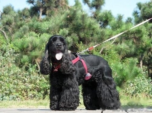
Americancockerdog
1. Canine congenital cataract: It begins in the embryonic period. Due to the abnormal development of the lens and its capsule in the mother, it manifests itself as a cataract after birth.
2. Traumatic cataract in dogs: due to various mechanical injuries (such as corneal penetration), the nutrition of the lens and its capsule is impaired.
3. Secondary cataract in dogs: secondary to other eye diseases and systemic diseases, such as iritis, uveitis, diabetes, etc.
4. Canine senile cataract: the degenerative changes of the lens, mainly seen in aged dogs aged 8-12.
:
1. Clinical symptoms
At present, the main basis for the diagnosis of cataract in dogs in China is the clinical diagnosis of the disease. Symptoms, when the dog has symptoms of vision loss, such as stumbling, often bumping into objects when walking in an unfamiliar environment, not seeing a thrown ball, strabismus, being easily frightened, being unusually sensitive to surrounding sounds, and having a tendency to be aggressive towards strangers. When you turn a blind eye to the danger, etc., you can suspect a cataract.
2. Laboratory examinations
After pupil dilation with 10 g/L tropicamide, the dog was brought into the dark room by an assistant and carried out Baoding, followed by a veterinary examination with a slit lamp or ophthalmoscope. If there is no ophthalmoscope and other equipment in the laboratory, a small flashlight and a magnifying glass can also be used to form a simple device to check [5].
Examination by the thorough imaging method, if there is a little shadow around the lens or the posterior pole, it can be suspected.
Slit-lamp microscope examination, the lens peripheral or posterior pole is pointy opacity, the cortex is still transparent; or the posterior pole of the lens has water scar-like changes, it can be judged as cataract.
3. Ultrasound examination
The ophthalmic ultrasound system can clearly distinguish various structures in the eye, and the image is clear and the effect is very good. Generally, a small ultrasonic probe above 7.5 MHz is required for cataract inspection. For large dogs, a 5 MHz probe is required to inspect the retro-ocular structure. If a clearer image is required, a higher frequency probe is required. When the dog suffers from eye pain or the animal is extremely uncooperative, sedation or local anesthesia or even general anesthesia is required. The normal canine lens only has obvious echoes in the anterior and posterior capsules of the lens on B-ultrasound. When the peripheral part of the lens has obvious weak or strong echoes, it can be judged that the dog has cataract.
Canine cataract is a disease that no medicine can cure, the color change of crystal is like the process of hard-boiled egg, from egg white to white. The only way to restore vision is to surgically remove the cloudy lens.
Before surgery, any rapidly growing cataract can cause eye irritation. Uveitis (inflammation of the inside of the eye) is usually caused by an abnormality of the lens. Whether cataract surgery is performed or not, uveitis needs prompt treatment, otherwise it will cause other problems of the eye.
Canine cataract surgery is the same as human cataract surgery. We call it phacoemulsification. During surgery, we dilate the pupil first, and then make a small opening at the edge of the cornea. Inside the eye, the lens is actually in a pocket, which we call a capsular bag. A small hole is torn in the capsular bag. , we put the ultrasound probe in and break the crystal, but not the capsular bag. After the lens is aspirated, we will place an intraocular lens. The intraocular lens has two loops that can be supported within the capsular bag. Finally, the corneal incision is sutured.
Post-operative care of canine cataracts is very important, and if post-operative care is neglected, it may adversely affect an otherwise successful surgery. Therefore, at this stage, the owner must take good care of the sick dog to avoid the dog being overexcited. It is very important to give the dog eye drops, wear an Elizabethan ring, and regularly review it.
Medical management: Use steroid anti-inflammatory drugs to speed up the healing of wound sutures, but when corneal ulcers occur, steroid drugs should be stopped immediately until the ulcers heal before continuing use; postoperative broad-spectrum should be used. Antibiotics to prevent bacterial infection; topical mydriatics (eg, atropine, tropicamide) for post-surgical uveitis to speed up the outflow of aqueous humor and reduce eye swelling.
Due to the lack of understanding and attention to canine cataract in the field of veterinary medicine, most of the affected dogs fail to receive timely treatment and become blind. I hope the materials provided in this article can help you.
![[Dog Training 5] The training method of pet dog dining etiquette](/static/img/12192/12192_1.jpg)


