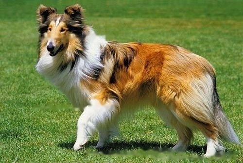
The Collie Sheepdog or Scottish Sheepdog is a flexible, sturdy, firm-standing dog with an active, lively personality. The broad and thick chest of the dog shows strength, the sloping shoulder blades and moderately curved hocks show speed and grace, and the face shows a very high IQ. The overall structure between the various parts of the dog's body is perfect and the proportions are harmonious. But like many dogs, it has many genetic defects. What are its genetic defects?

Scottish Sheepdog
1. Eye deformities
Curry The eye deformity of is mainly manifested as incomplete development of the eye. This disease has been reported worldwide, regardless of coarse or fine hair. This defect is manifested in the incomplete development of the choroid, optic disc or the area connected to it (coloboma). ), thinning of certain areas of the sclera, and retinal detachment. When these deformities occur, they are clinically manifested as reduced vision or complete blindness. Most Collie dogs with eye deformities do not show abnormal eye vision.
2. Permanent pupillary membrane
In the embryonic stage, the iris first forms a hard-nucleated mesoderm. Pupils atrophy in the later stages of pregnancy. In some animals, if these mesoderm filaments are found, we call them permanent pupillary membranes. These filaments are often seen in puppies of 6-8 weeks of age. But beyond the neonatal period is considered a defect.
In puppies 6-8 weeks, if these filaments are very large or present for more than 12 weeks, this should be noted in the record book, and this disease is common in Collies.
3. Progressive retinal atrophy
Progressive retinal atrophy is used to describe a hereditary retinal disease. Characterized by hypoplasia of cones and rods or progressive retinal atrophy. In the Collie, hypoplasia refers to a specific tissue in which both cones and rods are hypoplastic.
Affected animals are clinically symptomatic of night blindness at 12 weeks of age and then completely blind by about one year of age. All affected animals eventually became blind, and changes in the retina were observable at 6 months of age. The final stage was characterized by thinning of retinal blood vessels and hypertrophic choroidal carpet reflexes. Pigmentation changes in non-choroidal blanket reflex areas, pale optic disc.
Retinal potential map can be accurately diagnosed at 10-12 weeks of age. Retinal potential map is used to stimulate the eye with light and observe the changes in the internal potential of the eye, so as to judge the function of the retina. The distinction between cone and rod cell function by light stimulation can be performed before clinical symptoms appear.
There are hereditary defects, but they only show us the good side. We should take more care of the Collie.
![[Dog Training 5] The training method of pet dog dining etiquette](/static/img/12192/12192_1.jpg)


